Iliotibial Band Friction Syndrome - MSK Radiology Imaging

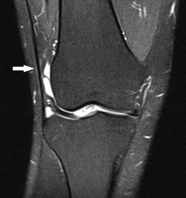
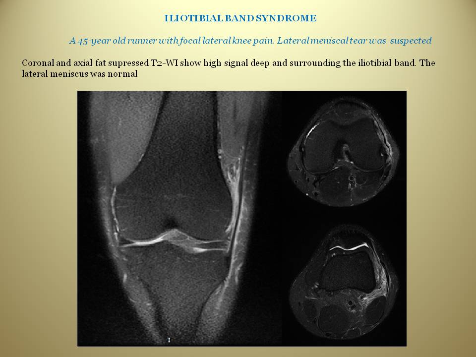
Iliotibial Band Friction Syndrome - MSK Radiology Imaging Findings: • The iliotibial band (ITB) is thickened with no tear visualized. • Ill-defined area of increased signal on fluid-sensitive sequences between the lateral femoral condyle (LFC) and ITB. Case description: • Clinical: anterolateral knee pain with point tenderness 1-2 cm proximal to lateral joint line. • Treatment: Conservative measures and image-guided steroid injection (may accelerate recovery). • Chronic inflammatory response to friction between the TIB and LFC causing ill-defined increased signal in this region on fluid-sensitive sequences. • Findings of chronic disease: - Thickening of the IT B and superficial increased T2-signal. - Reactive marrow edema in the adjacent LFC. Differential diagnosis for similar location of pain: • Fluid in lateral knee joint recess: Well-defined margins and connection to knee joint is seen. • Lateral collateral ligament complex injury: Signal around and/or within lateral ligaments. • Direct trauma/contusion: Soft-tissue swelling is predominant, with minimal fluid-signal deep to ITB. Dr. Donald von Borstel @DrvonBorstel #Iliotibial #Band #ITBand #Friction #Syndrome #Radiology #diagnosis #msk #clinical

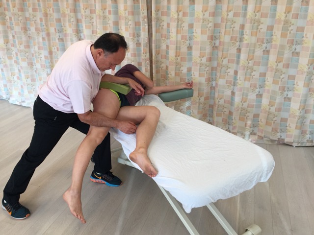
Ultrasound Guided IT Band Injection - Sports Medicine Review

The iliotibial tract: imaging, anatomy, injuries, and other pathology

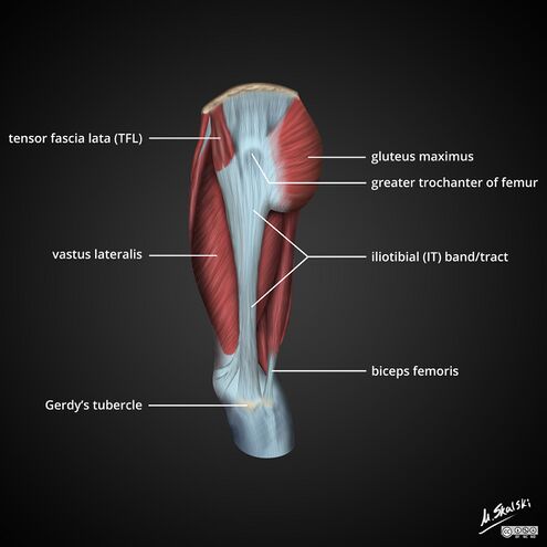
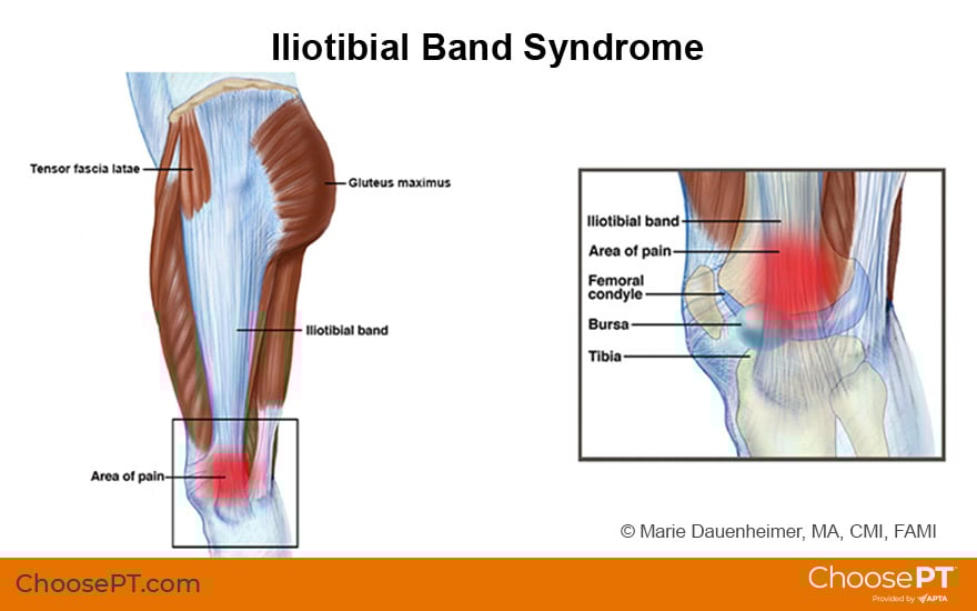
ITB ANATOMY, SYNDROME & TRAUMA, ILIOTIBIAL BAND MRI ANATOMY, SYNDROME, TRAUMA Finding and assessing the Iliotibial Band (ITB) on MRI of the Knee is the first structure we assess when

Ultrasound of iliotibial band syndrome

Do I need a knee x-ray for my knee pain?

Iliotibial band syndrome, Radiology Reference Article

Iliotibial band syndrome, Radiology Case
Iliotibial Tract - Physiopedia

EPOS™ - P-0075
EPOS™
Guide, Physical Therapy Guide to Iliotibial Band Syndrome (IT Band Syndrome)