Red Cell Staining (Color) • The Blood Project
Description
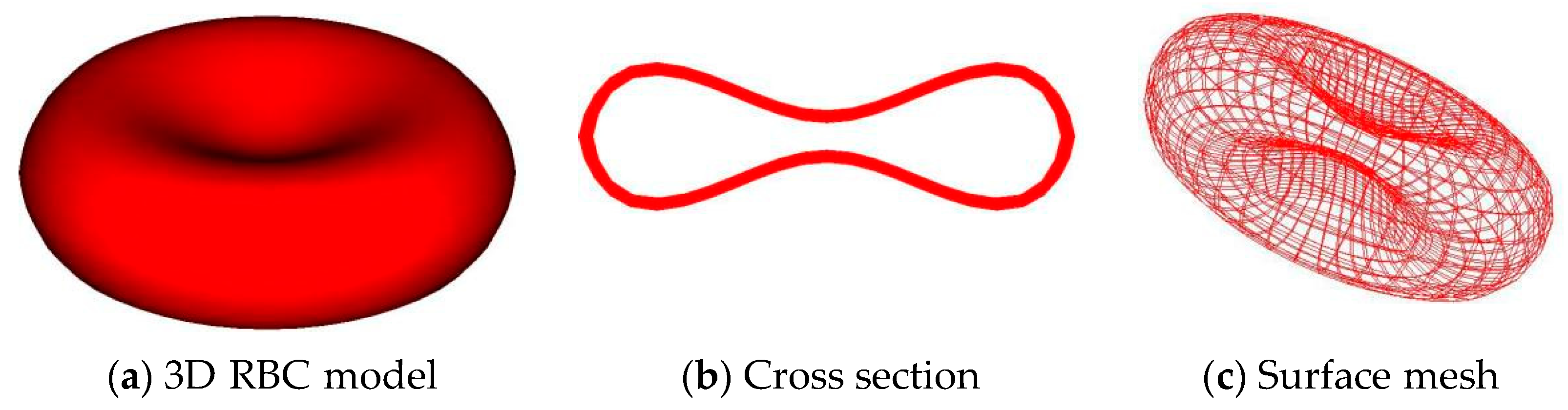
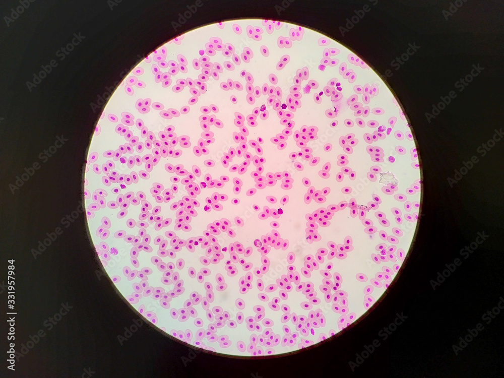
Central pallor Introduction A normal red blood cell has a biconcave disk shape. Because the center is much thinner than the periphery, it creates the

4,900+ Red Blood Cell Microscope Stock Photos, Pictures & Royalty
Red blood cell - Wikipedia
The Reason for Staining a Specimen on the Microscope
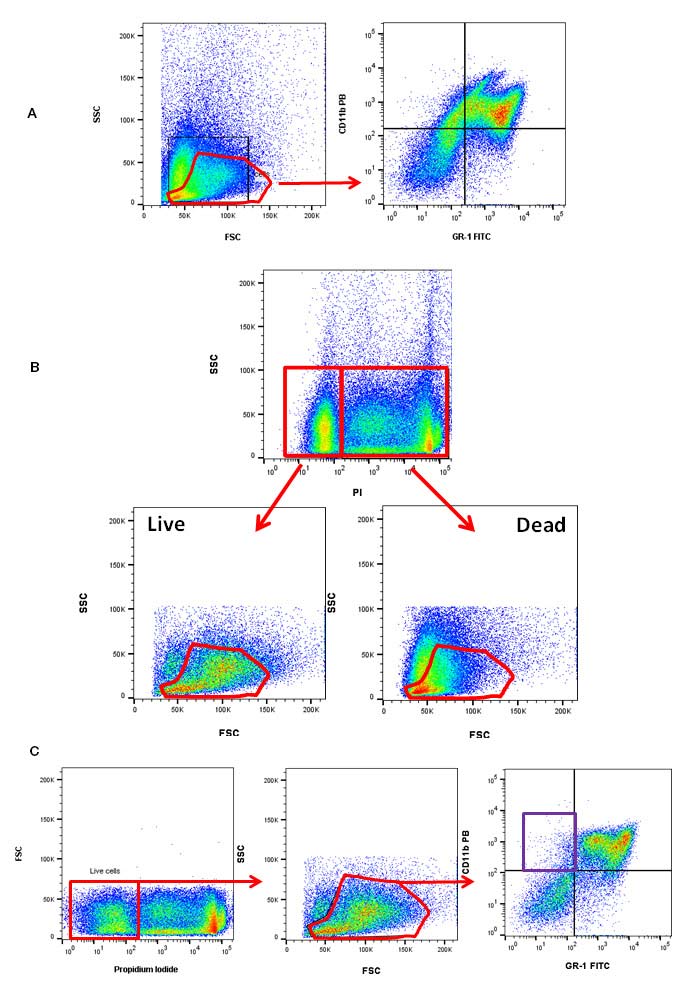
Controls for Flow Cytometry
Red blood cell of frog was dyed by Wright's stain. The red blood

H&E stain - Wikipedia
/images/vimeo_thumbnails/257904024/BzdbAdZUs4jZVZsV5e1ZNQ_overlay.jpg)
Erythrocytes - Histology, Structure, Function, Life Cycle

Morphology of red blood cells stained on day a) 0, b) 5, c) 10, d
Blood & Bodily Fluids - Forensic Resources

File:Human red blood cell.jpg - Wikimedia Commons
Automated cell counting in IHC-DAB
Related products


$ 32.50USD
Score 4.8(390)
In stock
Continue to book
$ 32.50USD
Score 4.8(390)
In stock
Continue to book
©2018-2024, farmersprotest.de, Inc. or its affiliates