The integration of artificial intelligence with contrast-enhanced mammogram in the work up of suspicious breast lesions: what do you expect?, Egyptian Journal of Radiology and Nuclear Medicine
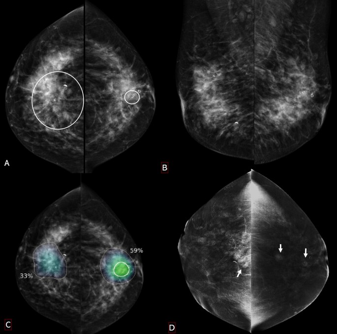
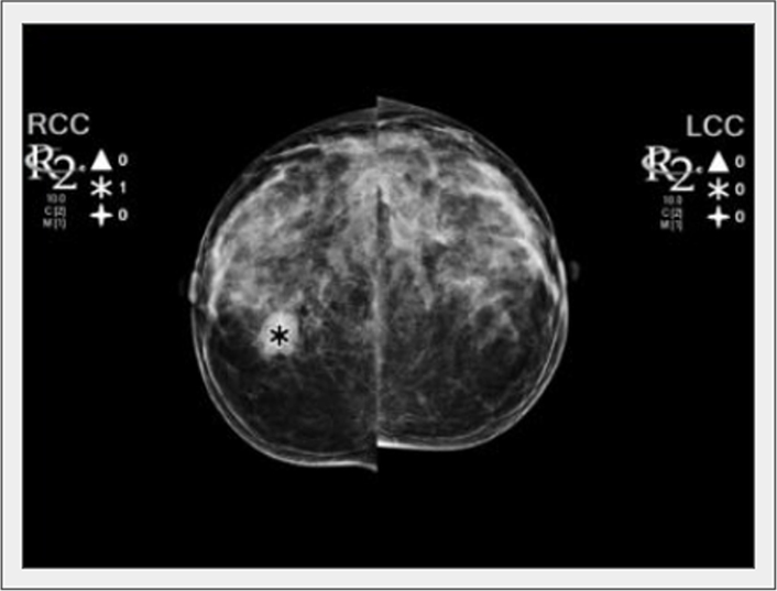
The enhancement overlaps at contrast-enhanced mammogram (CEM) between benign and malignant breast abnormalities presents a high probability of false-positive lesions and subjects females’ candidate for screening and diagnostic mammograms to unnecessary biopsy and anxiety. The current work aimed to evaluate the ability of mammograms scanned by artificial intelligence (AI) to enhance the specificity of CEM and support the probability of malignancy in suspicious and malignant looking breast lesions. The study included 1524 breast lesions. The AI algorithm applied to the initial mammograms and generated location information for lesions. AI scoring suggested the probability of malignancy ranged from 100% (definite cancers) and < 10% (definite non-cancer) and correlated with recombinant contrast enhanced images. The malignant proved abnormalities were 1165 (76.5%), and the benign ones were 359 (26.5%). BI-RADS 4 category was assigned in 704 lesions (46.2%) divided into 400 malignant (400/704, 56.8%) and 304 benign (304/704, 43.2%). BI-RADS 5 category presented by 820 lesions (53.8%), 765 of them were malignant (765/820, 93.3%) and 55 were benign (55/820, 6.7%). The sensitivity of digital mammogram whether supported by AI (93.9%) or contrast media (94.4%) was significantly increased to 97.2% (p < 0.001) when supported by both methods. Improvement of the negative predictive value (from 80.6% and 79.6% to 89.8%, p < 0.05) and the accuracy (from 91.1 and 88.8 to 94.0%, p < 0.01) was detected. Contrast-enhanced mammogram helps in specification of different breast lesions in view of patterns of contrast uptake and morphology descriptors, yet with some overlap. The use of artificial intelligence applied on digital mammogram reduced the interpretational variability and limited attempts of re-biopsies of suspicious looking breast lesions assessed by contrast-enhanced mammograms.

The integration of artificial intelligence with contrast-enhanced mammogram in the work up of suspicious breast lesions: what do you expect?, Egyptian Journal of Radiology and Nuclear Medicine
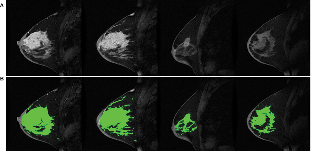
Frontiers Current and Emerging Magnetic Resonance-Based Techniques for Breast Cancer

Artificial intelligence for breast cancer detection in mammography and digital breast tomosynthesis: State of the art - ScienceDirect

A review on machine learning techniques for the assessment of image grading in breast mammogram

The integration of artificial intelligence with contrast-enhanced mammogram in the work up of suspicious breast lesions: what do you expect?, Egyptian Journal of Radiology and Nuclear Medicine
PDF) Comparing the Diagnostic Performance of Contrast-Enhanced Mammography and Breast MRI: a Systematic Review and Meta-Analysis

Does artificial intelligence aid in the detection of different types of breast cancer?, Egyptian Journal of Radiology and Nuclear Medicine

Artificial intelligence for breast cancer detection in mammography and digital breast tomosynthesis: State of the art - ScienceDirect

Diagnostics, Free Full-Text
PDF) Interpretation of patterns of enhancement on contrast-enhanced spectral mammography: An approach to a standardized scheme

Recent advancements in artificial intelligence for breast cancer: Image augmentation, segmentation, diagnosis, and prognosis approaches - ScienceDirect

Performance of AI-aided mammography in breast cancer diagnosis: Does breast density matter?, Egyptian Journal of Radiology and Nuclear Medicine

Calcifications at Digital Breast Tomosynthesis: Imaging Features and Biopsy Techniques
Artificial intelligence in breast cancer imaging: risk stratification, lesion detection and classification, treatment planning and prognosisa narrative review