Standing anteroposterior and lateral X-rays of the dorso-lumbar spine

Download scientific diagram | Standing anteroposterior and lateral X-rays of the dorso-lumbar spine showing a failure of the pedicular screws at T11. Note the iatrogenic flat-back deformity with loss of sagittal spine alignment and +ve sagittal vertical axis. from publication: Acute Paraplegia Secondary to Thoracic Disc Herniation of the Adjacent Segment Following Thoracolumbar Fusion and Instrumentation | Proximal junctional disease is a well-recognized postoperative phenomenon in adults who are undergoing long thoracolumbar fusion and instrumentation, and is attributed to increased a junctional stress concentration. In general, the onset of symptoms in these patients is | Paraplegia, Fusion and Segmentation | ResearchGate, the professional network for scientists.

Standing anteroposterior and lateral radiographs of the lumbar spine
Xray cervical spine AP and lateral shows straightening of cervical spine? What does it mean? - Quora

Frontiers Case Report: Campylobacter fetus caused pyogenic spondylodiscitis with a presentation of cauda equina syndrome after instrumented lumbar fusion surgery

/publication/359342356/figure/
X-Ray of the Spine
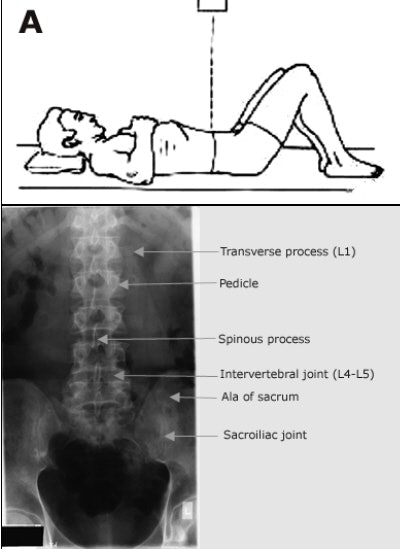
The lowdown on lumbar spine positioning

Thoracic spine (lateral view), Radiology Reference Article

MedPix Case - Cervical and Lumbar Radiculopathy Complicated by Knee and Shoulder Injury.

A Case Series That Supports the Application of the S2AI Technique for Fractures and Failures After Lumbosacral Fusion