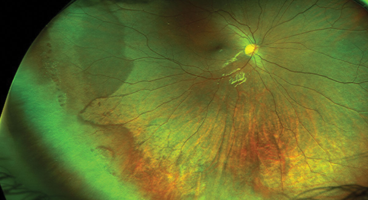
Ultra-wide-field fundus photographs and ultra-wide-field

Download scientific diagram | Ultra-wide-field fundus photographs and ultra-wide-field fluorescein angiographic imaging of ocular toxocariasis. (A) A granuloma with mild vitreous opacity. (B) A tractional retinal fold with localized tractional retinal detachment. (C) Diffuse peripheral vascular leakage. (D) A prominent optic disc leakage. from publication: The Clinical Characteristics of Ocular Toxocariasis in Jeju Island Using Ultra-wide-field Fundus Photography | Toxocariasis, Ocular and Photography | ResearchGate, the professional network for scientists.

Ultrawide-field color fundus photography (1) and ultrawide-field fundus
Ultra-widefield Imaging Ideal for Monitoring Myopic Maculopathy

PDF) The Clinical Characteristics of Ocular Toxocariasis in Jeju Island Using Ultra-wide-field Fundus Photography
Ultra-wide-field fundus photographs and ultra-wide-field fluorescein

Ultra-wide field fundus photography (AeC) and fluorescein angiography

Ultra-wide-field fundus photographic findings of a patient with ocular

Jong Young Lee's research works Jeju National University Hospital, Jeju City and other places

Life, Free Full-Text
The Clinical Utility of Ultra-Wide-Field Imaging
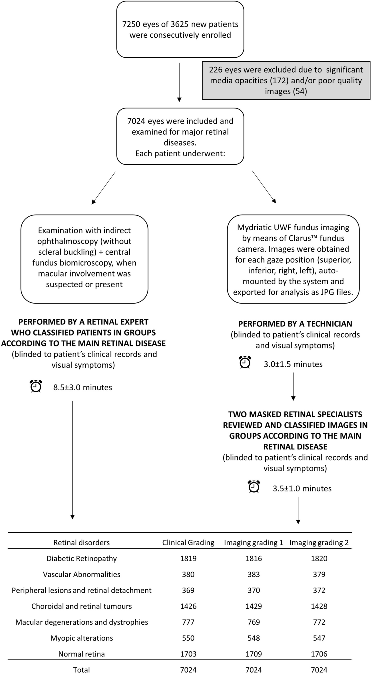
Ultra-wide-field fundus photography compared to ophthalmoscopy in diagnosing and classifying major retinal diseases